 これらの AFM プローブ は他のお好きなBudgetSensors AFM プローブ と同時に購入してBudget Combo Boxを作ることができます!
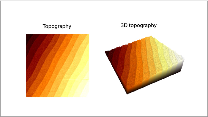
SrTiO3単結晶基板の表面形状像とその3D表示
スキャン BudgetSensors ContAl-G AFMプローブ コンタクトモード 5umスキャン
Image courtesy of Prof. Yunseok Kim Sungkyunkwan University, South Korea
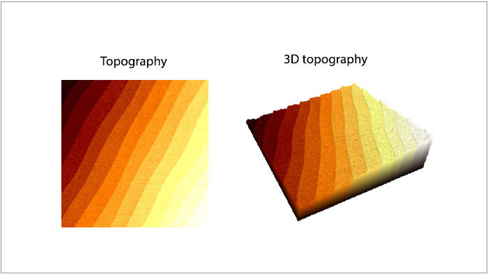
多結晶SrTiO3 単結晶基板の表面形状とその3D表示
スキャン BudgetSensors ContAl-G AFM プローブ コンタクトモード 5umスキャン
Image courtesy of Prof. Yunseok Kim, Sungkyunkwan University, South Korea
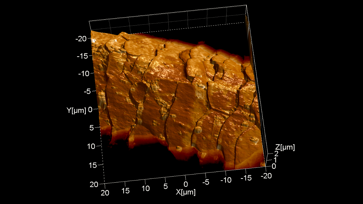
コンタクトモードの形状情報を3Dで表示し、ラテラルフォースモードの情報をオーバーレイした。うろこ状のモルフォロジーとヘアスプレーの液滴を観察できた。
スキャン BudgetSensors ContAl-G AFM プローブ, 40 umスキャン
Image courtesy of Dr. Yordan Stefanov, Innovative Solutions Bulgaria
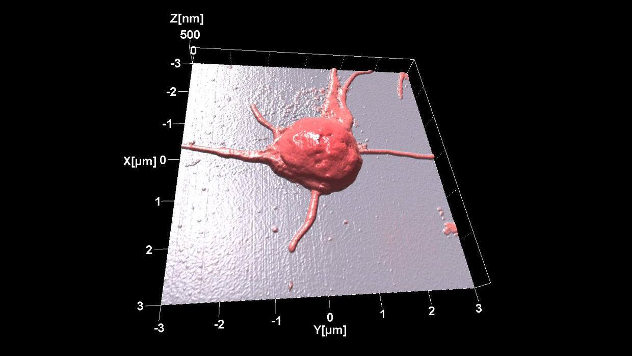
室温で取得したヒト血小板の形状情報
スキャン BudgetSensors ContAl-G AFM プローブ, 6 umスキャン
Image courtesy of Dr. Tonya Andreeva, Institute of Biophysics and Biomedical Engineering, BAS
